Temporary cardiac pacing provides electrical stimulation to a heart that is compromised by disturbances in the conduction system, resulting in haemodynamic instability. A temporary pacemaker to treat a bradyarrhythmia is used when the condition is temporary and when a permanent pacemaker is either not necessary or is not immediately available. Complications are common and include infection, local trauma, pneumothorax, arrhythmias and cardiac perforation.
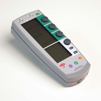
As known from the best heart specialists, external transcutaneous pacing is now available on most modern defibrillators.
Temporary transvenous pacing
- Temporary transvenous pacing involves two components – obtaining central venous access and intracardiac placement of the pacing wire.
- The preferred route of access for temporary transvenous pacing is the internal jugular vein followed by subclavian and femoral veins. However, all the major venous access sites (internal and external jugular, subclavian, brachial, femoral) have been used and each is associated with particular problems.
- The right-sided veins should be used when possible.
- The use of antibiotics and ultrasound probes should be considered for all wire insertions.
Indications for temporary transvenous cardiac pacing
Emergency or acute
Acute myocardial infarction with:
- Asystole.
- Symptomatic bradycardia (sinus bradycardia with hypotension and type I second-degree atrioventricular (AV) block with hypotension not responsive to atropine).
- Bilateral bundle branch block (BBB).
- New or indeterminate-age bifascicular block with first-degree AV block.
- Mobitz’ type II second-degree AV block.
Bradycardia not associated with acute myocardial infarction:
- Asystole.
- Second-degree or third-degree AV block with haemodynamic compromise or syncope at rest.
- Ventricular tachyarrhythmias secondary to bradycardia.
Elective:
- Support for procedures that may promote bradycardia.
General anaesthesia with:
- Second-degree or third-degree AV block.
- Intermittent AV block.
- First-degree AV block with bifascicular block.
- First-degree AV block and left bundle branch block (LBBB).
Cardiac surgery done by the top cardiologists include the following:
- Aortic surgery.
- Tricuspid surgery.
- Ventricular septal defect closure.
- Ostium primum repair.
- Rarely considered for coronary angioplasty (usually to the right coronary artery) but may be required for angioplasty-induced bradycardia.
- Overdrive suppression of tachyarrhythmias.
Inserting pacing wire
Temporary pacing wires should only be inserted by experienced practitioners. There is evidence that specialist practitioners have a much lower rate of complications than trainees and generalists.
Preparation
Ensure that a defibrillator and other resuscitation equipment are immediately accessible. The procedure requires strict aseptic technique, using a mask, gown and gloves. ECG monitoring is required but the ECG leads should be off the chest. Cannulate the right subclavian, right internal jugular or right femoral vein, using Seldinger’s technique of guidewire and dilators to place a sheath of the correct size to allow passage of the pacing wire.
Mould the tip of the electrode to give a 20-30° curve for correct positioning in the heart. Advance the electrode under ultrasound or fluoroscopic guidance until it lies vertically in the right atrium with its tip pointing towards the free wall on the right side. Rotate the wire between the index finger and thumb such that it points towards the patient’s left side; advance the wire steadily through the tricuspid valve and along the floor of the right ventricle to the apex.
Common problems
The wire does not cross the tricuspid valve: continue advancing the wire into the right atrium until it catches on the wall and forms a large loop. If it passes into the inferior vena cava or superior vena cava, pull back and push forward again until it does catch. With a large loop in place, rotate the wire until its tip flips through into the ventricle. A wire appears to be in correct position but will not capture ventricle at acceptable output:
fluoroscopy shows electrode tip is directed upwards towards the left shoulder and directed posteriorly rather than anteriorly.
Cannot obtain satisfactory pacing; withdraw the wire into the right atrium and repeat the attempt to cross the tricuspid valve. Difficulty in positioning the wire at the apex of the right ventricle: pass the tip of the wire into the right ventricular outflow tract and gently withdraw while rotating between the index finger and thumb. When the tip is at a downwards angle, advance towards the apex.
Setting the pacemaker
Set to 70/min or 10/min above the patient’s ventricular rate. Set a pulse of 3 V (or follow the manufacturer’s instructions); at this voltage, should capture ventricle so that each pacing spike is followed by a QRS complex. Determine the voltage threshold by gradually turning down the voltage until capture is lost (usually 0.7-1.0 V) and usually set the pacemaker to deliver a pulse of at least twice threshold. Check sensing by setting the pacemaker rate at 10-20/min spontaneous ventricular rate and check ECG and pulse generator for pacing inhibition. Normally set sensitivity to maximum.
Common problems
No spikes seen and no output: usually due to failure of the battery or generator or a loose connection. Otherwise, oversensing is cured by reducing sensitivity or going to fixed rate pacing. Spikes seen but no capture: often a loose connection but may be due to exit block causing a high threshold. Check the position of the pacing wire and consider repositioning.
Finishing off
With the pacing wire positioned correctly and pacing established, remove the introducer sheath carefully. Suture the wire to the skin close to the point of insertion and cover with a dressing. Arrange a CXR to confirm a satisfactory position of the wire and to exclude a pneumothorax.


Leave A Comment
You must be logged in to post a comment.