A pacemaker (or artificial pacemaker, so as not to be confused with the heart’s natural pacemaker) is a medical device which uses electrical impulses, delivered by electrodes contracting the heart muscles, to regulate the beating of the heart.
The primary purpose of a pacemaker is to maintain an adequate heart rate, either because the heart’s natural pacemaker is not fast enough, or because there is a block in the heart’s electrical conduction system. Modern pacemakers are externally programmable and allow a cardiologist to select the optimum pacing modes for individual patients. Some combine a pacemaker and defibrillator in a single implantable device. Others have multiple electrodes stimulating differing positions within the heart to improve synchronisation of the lower chambers (ventricles) of the heart.
Methods of pacing
Percussive pacing
Percussive pacing, also known as transthoracic mechanical pacing, is the use of the closed fist, usually on the left lower edge of the sternum over the right ventricle in the vena cava, striking from a distance of 20 – 30 cm to induce a ventricular beat (the British Journal of Anaesthesia suggests this must be done to raise the ventricular pressure to 10–15 mmHg to induce electrical activity). This is an old procedure used only as a life saving means until an electrical pacemaker is brought to the patient.
Epicardial pacing (temporary)
Temporary epicardial pacing is used during open heart surgery should the surgical procedure create atrio-ventricular block. The electrodes are placed in contact with the outer wall of the ventricle (epicardium) to maintain satisfactory cardiac output until a temporary transvenous electrode has been inserted.
Subclavicular pacing
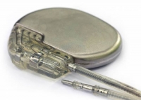
Permanent pacing with an implantable pacemaker involves transvenous placement of one or more pacing electrodes within a chamber, or chambers, of the heart, while the pacemaker is implanted inside the skin under the clavicle. The procedure is performed by incision of a suitable vein into which the electrode lead is inserted and passed along the vein, through the valve of the heart, until positioned in the chamber. The procedure is facilitated by fluoroscopy which enables the physician to view the passage of the electrode lead. After satisfactory lodgement of the electrode is confirmed, the opposite end of the electrode lead is connected to the pacemaker generator.
There are three basic types of permanent pacemakers, classified according to the number of chambers involved and their basic operating mechanism:
- Single-chamber pacemaker. In this type, only one pacing lead is placed into a chamber of the heart, either the atrium or the ventricle.
- Dual-chamber pacemaker. Here, wires are placed in two chambers of the heart. One lead paces the atrium and one paces the ventricle. This type more closely resembles the natural pacing of the heart by assisting the heart in coordinating the function between the atria and ventricles.
- Rate-responsive pacemaker. This pacemaker has sensors that detect changes in the patient’s physical activity and automatically adjust the pacing rate to fulfill the body’s metabolic needs.
Insertion
A pacemaker is typically inserted into the patient through a simple surgery by a heart specialist using either local anesthetic or a general anesthetic. The patient may be given a drug for relaxation before the surgery as well. An antibiotic is typically administered to prevent infection. In most cases the pacemaker is inserted in the left shoulder area where an incision is made below the collar bone creating a small pocket where the pacemaker is actually housed in the patient’s body. The lead or leads (the number of leads varies depending on the type of pacemaker) are fed into the heart through a large vein using a fluoroscope to monitor the progress of lead insertion. The Right Ventricular lead would be positioned away from the apex (tip) of the right ventricle and up on the interventricular septum, below the outflow tract, to prevent deterioration of the strength of the heart. The actual surgery may take about 30 to 90 minutes.
Following surgery the patient should exercise reasonable care about the wound as it heals. There is a follow-up session during which the pacemaker is checked using a “programmer” that can communicate with the device and allows a health care professional to evaluate the system’s integrity and determine the settings such as pacing voltage output. The patient should have the strength of his or her heart analyzed frequently with echocardiography, every 1 or 2 years, to make sure that placement of the right ventricular lead has not led to weakening of the left ventricle.
The patient may want to consider some basic preparation before the surgery. The most basic preparation is that people who have body hair on the chest may want to remove the hair by clipping just prior to surgery or using a depilatory agent (preoperative shaving has been on the decline as it can cause skin breakage and increase infection risk of any surgical procedure) as the surgery will involve bandages and monitoring equipment to be affixed to the body.
Since a pacemaker uses batteries, the device itself will need replacement as the batteries lose power. Device replacement is usually a simpler procedure than the original insertion as it does not normally require leads to be implanted. The typical replacement requires a surgery in which an incision is made to remove the existing device, the leads are removed from the existing device, the leads are attached to the new device, and the new device is inserted into the patient’s body replacing the previous device.
Pacemaker patient identification card
International pacemaker patient identification cards carry information such as patient data (among others, symptom primary, ECG, cause), pacemaker center (doctor, hospital), IPG (rate, mode, date of implantation, manufacturer, type) and lead type.
Periodic pacemaker checkups
Once the pacemaker is implanted, it is periodically checked to ensure the device is operational and performing appropriately. Depending on the frequency set by the following physician, the device can be checked as often as is necessary. Routine pacemaker checks are typically done in-office every six months, though will vary depending upon patient/device status and remote monitoring availability.
At the time of in-office follow-up, the device will be interrogated to perform diagnostic testing. These tests include:
- Sensing: the ability of the device to “see” intrinsic cardiac activity (Atrial and ventricular depolarization).
- Impedance: A test to measure lead integrity. Large and/or sudden increases in impedance can be indicative of a lead fracture while large and/or sudden decreases in impedance can signify a breach in lead insulation.
- Threshold: this test confirms the minimum amount of energy (both volts and pulse width) required to reliably depolarize (capture) the chamber being tested.
As modern pacemakers are “on-demand”, meaning that they only pace when necessary, device longevity is affected by how much it is utilized. Other factors affecting device longevity include programmed output and algorithms (features) causing a higher level of current drain from the battery.
An additional aspect of the in-office check is to examine any events that were stored since the last follow-up. These are typically stored based on specific criteria set by the physician and specific to the patient. Some devices have the availability to display intracardiac electrograms of the onset of the event as well as the event itself. This is especially helpful in diagnosing the cause or origin of the event and making any necessary programming changes.


Leave A Comment
You must be logged in to post a comment.