Tachycardia is a common type of heart rhythm disorder (arrhythmia) in which the heart beats faster than normal while at rest.
It’s normal for your heart rate to rise during exercise or as a physiological response to stress, trauma or illness (sinus tachycardia). But in tachycardia the heart beats faster than normal in the upper or lower chambers of the heart or both while at rest.
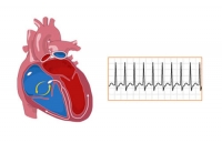
Your heart rate is controlled by electrical signals sent across heart tissues. Tachycardia occurs when an abnormality in the heart produces rapid electrical signals that quicken the heart rate, which is normally about 60 to 100 beats a minute at rest.
Symptoms
When your heart is beating too fast, it may not pump blood effectively to the rest of your body. This can deprive your organs and tissues of oxygen and can cause the following tachycardia-related signs and symptoms:
- Shortness of breath
- Lightheadedness
- Rapid pulse rate
- Heart palpitations — a racing, uncomfortable or irregular heartbeat or a sensation of “flopping” in the chest
- Chest pain
- Fainting (syncope)
Some people with tachycardia have no symptoms, and the condition is only discovered during a physical examination or with a heart-monitoring test called an electrocardiogram.
Causes
Tachycardia is caused by something that disrupts the normal electrical impulses that control the rate of your heart’s pumping action. Many things can cause or contribute to problems with the heart’s electrical system. These include:
- Damage to heart tissues from heart disease
- Abnormal electrical pathways in the heart present at birth (congenital heart conditions, including long QT syndrome)
- Disease or congenital abnormality of the heart
- Anemia
- Exercise
- Sudden stress, such as fright
- High or low blood pressure
- Smoking
- Fever
- Drinking too much alcohol
- Drinking too many caffeinated beverages
- Medication side effects
- Abuse of recreational drugs, such as cocaine
- Imbalance of electrolytes, mineral-related substances necessary for conducting electrical impulses
- Overactive thyroid (hyperthyroidism)
In some cases, the exact cause of tachycardia can’t be determined.
Risk factors
Any condition that puts a strain on the heart or damages heart tissue can increase your risk of tachycardia. Lifestyle changes or medical treatment may decrease the risk associated with the following factors:
- Heart disease
- High blood pressure
- Sleep apnea
- Overactive or underactive thyroid
- Smoking
- Diabetes
- Heavy alcohol use
- Heavy caffeine use
- Use of recreational drugs
- Psychological stress or anxiety
- Anemia
Diagnosis
To diagnose your condition and determine the specific type of tachycardia, your cardiologist will evaluate your symptoms, conduct a thorough physical examination, and ask you about your health habits and medical history.
Several heart tests also may be necessary to diagnose tachycardia.
Electrocardiogram (ECG)
An electrocardiogram, also called an ECG or EKG, is the most common tool used to diagnose tachycardia. It’s a painless test that detects and records your heart’s electrical activity using small sensors (electrodes) attached to your chest and arms.
An ECG records the timing and strength of electrical signals as they travel through your heart. Your heart specialist can look for patterns among these signals to determine what kind of tachycardia you have and how abnormalities in the heart may be contributing to a fast heart rate.
Your doctor may also ask you to use portable ECG devices at home to provide more information about your heart rate.
Holter monitor. This portable ECG device is carried in your pocket or worn on a belt or shoulder strap. It records your heart’s activity for an entire 24-hour period, which provides your doctor with a prolonged look at your heart rhythms.
Your doctor will likely ask you to keep a diary during the same 24 hours. You’ll describe any symptoms you experience and record the time they occur.
Event monitor. This portable ECG device is intended to monitor your heart activity over a few weeks to a few months. You wear it all day, but it records only at certain times for a few minutes at a time.
With many event monitors, you activate them by pushing a button when you experience symptoms of a fast heart rate. Other monitors automatically sense abnormal heart rhythms and then start recording. These monitors allow your doctor to look at your heart rhythm at the time of your symptoms.
Electrophysiological test
Your cardiologist may recommend an electrophysiological test to confirm the diagnosis or to pinpoint the location of problems in your heart’s circuitry.
During this test, a doctor inserts thin, flexible tubes (catheters) tipped with electrodes into your groin, arm or neck and guides them through your blood vessels to various spots in your heart. Once in place, the electrodes can precisely map the spread of electrical impulses during each beat and identify abnormalities in your circuitry.
Cardiac imaging
Imaging of the heart may be performed to determine if structural abnormalities are affecting blood flow and contributing to tachycardia.
Types of cardiac imaging used to diagnose tachycardia include:
Echocardiogram (echo). An echocardiogram creates a moving picture of your heart using sound waves. An echo can identify areas of poor blood flow, abnormal heart valves and heart muscle that’s not working normally.
Magnetic resonance imaging (MRI). A cardiac MRI can provide still or moving pictures of how the blood is flowing through the heart and detect irregularities.
Computerized tomography (CT). CT scans combine several X-ray images to provide a more detailed cross-sectional view of the heart.
Coronary angiogram. To study the flow of blood through your heart and blood vessels, your heart specialist may use a coronary angiogram to reveal potential blockages or abnormalities. It uses a dye and special X-rays to show the inside of your coronary arteries.
Chest X-ray. This test is used to take still pictures of your heart and lungs and can detect if your heart is enlarged.
Stress Test
Your doctor may recommend a stress test to see how your heart functions while it is working hard during exercise or when medication is given to make it beat fast.
In an exercise stress test, electrodes are placed on your chest to monitor heart function while you exercise, usually by walking on a treadmill. Other heart tests may also be performed in conjunction with a stress test.
Tilt table test
This test is sometimes used to help your cardiologist better understand how your tachycardia contributes to fainting spells. Under careful monitoring, you’ll receive a medication that causes a tachycardia episode.
You lie flat on a special table, and then the table is tilted as if you were standing up. Your doctor observes how your heart and nervous system respond to these changes in position.
Additional tests
Your doctor may order additional tests as needed to diagnose an underlying condition that is contributing to tachycardia and judge the condition of your heart.
Treatment
Treatments for tachycardia are designed to address the cause of the condition as well as slow a fast heart rate when it occurs, prevent future episodes and minimize complications.
Stopping a fast heart rate
A fast heartbeat may correct itself, and you may be able to slow your heart rate using simple physical movements. However, you may need medication or other medical treatment to slow down your heartbeat.
Ways to slow your heartbeat include:
Vagal maneuvers. Your heart specialist may ask you to perform an action, called a vagal maneuver, during an episode of a fast heartbeat.
Vagal maneuvers affect the vagus nerve, which helps regulate your heartbeat. The maneuvers include coughing, bearing down as if you’re having a bowel movement and putting an ice pack on your face.
Medications. If vagal maneuvers don’t stop the fast heartbeat, you may need an injection of an anti-arrhythmic medication to restore a normal heart rate. An injection of this drug is administered at a hospital.
Your doctor also may prescribe a pill version of an anti-arrhythmic drug to take if you have an episode of a fast heartbeat that doesn’t respond to vagal maneuvers.
Cardioversion. In this procedure, a shock is delivered to your heart through paddles, an automated external defibrillator (AED) or patches on your chest. The current affects the electrical impulses in your heart and restores a normal rhythm. It’s generally used when emergency care is needed or when maneuvers and medications aren’t effective.
Preventing episodes of a fast heart rate
With the following treatments, it may be possible to prevent or manage episodes of tachycardia.
Catheter ablation. This procedure is often used when an extra electrical pathway is responsible for an increased heart rate.
In this procedure, a doctor inserts catheters into your groin, arm or neck and guides them through the blood vessels to your heart. Electrodes at the catheter tips can use extreme cold or radiofrequency energy to damage (ablate) the extra electrical pathway and prevent it from sending electrical signals.
Catheter ablation does not require surgery to access the heart, but it may also be performed in conjunction with other heart valve or artery repair surgeries.
Medications. Pill versions of anti-arrhythmic medications may prevent a fast heart rate when taken regularly.
Other types of drugs, such as calcium channel blockers and beta blockers, may be prescribed either as an alternative to or in combination with anti-arrhythmic medications.
Pacemaker. Some types of tachycardias may be treated with a pacemaker. A pacemaker is a small device that’s surgically implanted under your skin. When the device senses an abnormal heartbeat, it emits an electrical pulse that helps the heart resume a normal beat.
Implantable cardioverter. If you’re at risk of having a life-threatening tachycardia episode, your doctor may recommend an implantable cardioverter-defibrillator (ICD).
The pager-sized device is surgically implanted in your chest. The ICD continuously monitors your heartbeat, detects an increase in heart rate and delivers precisely calibrated electrical shocks, if needed, to restore a normal heart rhythm.
Surgery. Open-heart surgery may be needed in some cases to destroy an extra electrical pathway causing tachycardia.
In another type of surgery, called the maze procedure, a surgeon makes small incisions in heart tissue to create a pattern or maze of scar tissue. Because scar tissue doesn’t conduct electricity, it interferes with stray electrical impulses that cause some types of tachycardia.
Surgery is usually used only when other treatment options don’t work or when surgery is needed to treat another heart disorder.
Preventing blood clots
Some people with tachycardia have an increased risk of developing a blood clot that could cause a stroke or heart attack. Your cardiologist may prescribe a blood-thinning medication to help lower your risk.
Treating an underlying disease
If another medical condition is contributing to tachycardia, such as some form of heart disease or hyperthyroidism, treating the underlying problem may prevent or minimize tachycardia episodes.


Leave A Comment
You must be logged in to post a comment.